NanoEcho · Next-Level Diagnostic Imaging

See inside the body in real-time 3D.

A clinical-grade diagnostic platform that turns conventional ultrasound into a 3D, AI-highlighted view of what's actually happening inside the patient — tumours, infections, abnormalities — all revealed earlier and more clearly.

3D
Real-time render
AI
Highlighted findings
IoT
Connected device
3
Tier architecture
NanoEcho — next level diagnostics
Why we built it

Conventional ultrasound is fast — but flat.

Radiologists work from 2D black-and-white slices. Small lesions, early-stage infections and subtle abnormalities can be missed. Diagnoses get delayed. Patients get re-scanned. Treatment windows narrow.

NanoEcho was built to change that — by giving clinicians a live, 3D, AI-highlighted view of the anatomy in front of them, with the regions of concern called out automatically.

See more, sooner

Volumetric 3D reveals what flat slices hide — small lesions and infections detected earlier in the diagnostic journey.

AI points to the answer

Computer-vision overlays automatically highlight regions of concern, so the radiologist's eye goes to the right place first.

Faster, more confident calls

Clearer imagery and pre-annotated findings shorten reporting time and reduce the need for repeat scans.

How it works

From probe to 3D, in real time.

Four coordinated steps turn raw ultrasonic signal into a diagnostic-grade 3D image.

01

Scan

A high-precision ultrasonic probe streams live data from inside the body — slice by slice — at clinical frame-rates.

02

Excite

A controlled magnetic field excites administered nano-particles, making infections, tumours and abnormalities far more visible to the probe.

03

Enhance

AI computer-vision sharpens the imagery, reduces noise and highlights areas of concern for the radiologist to review first.

04

Render

A real-time 3D engine reconstructs the volume so the clinician can rotate, slice and explore the anatomy — not just look at a frame.

The clinician's experience

A purpose-built interface for the imaging room.

Calm, dark, glanceable — designed for a quiet clinical environment, with alarm + system-ready states always visible.

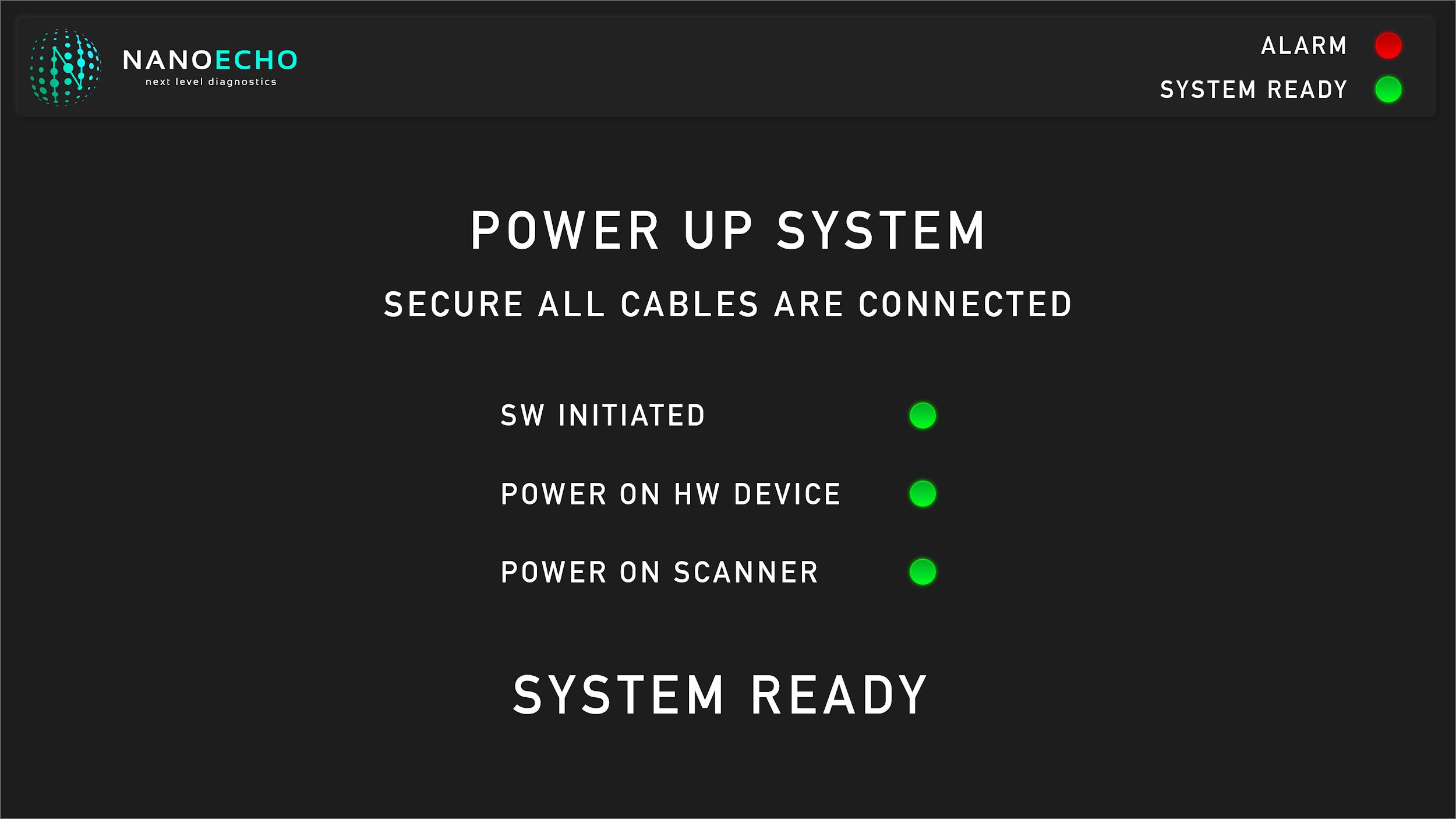
System Ready startup screen
System Ready

Pre-flight check confirms software, hardware and scanner power-on before a scan can start.

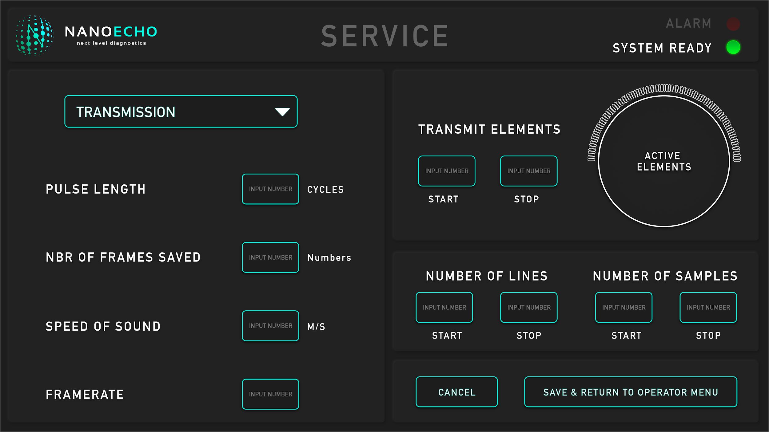
Service configuration screen
Service Configuration

Service-mode controls — pulse length, framerate, transmit elements — for biomedical engineers, not clinicians.

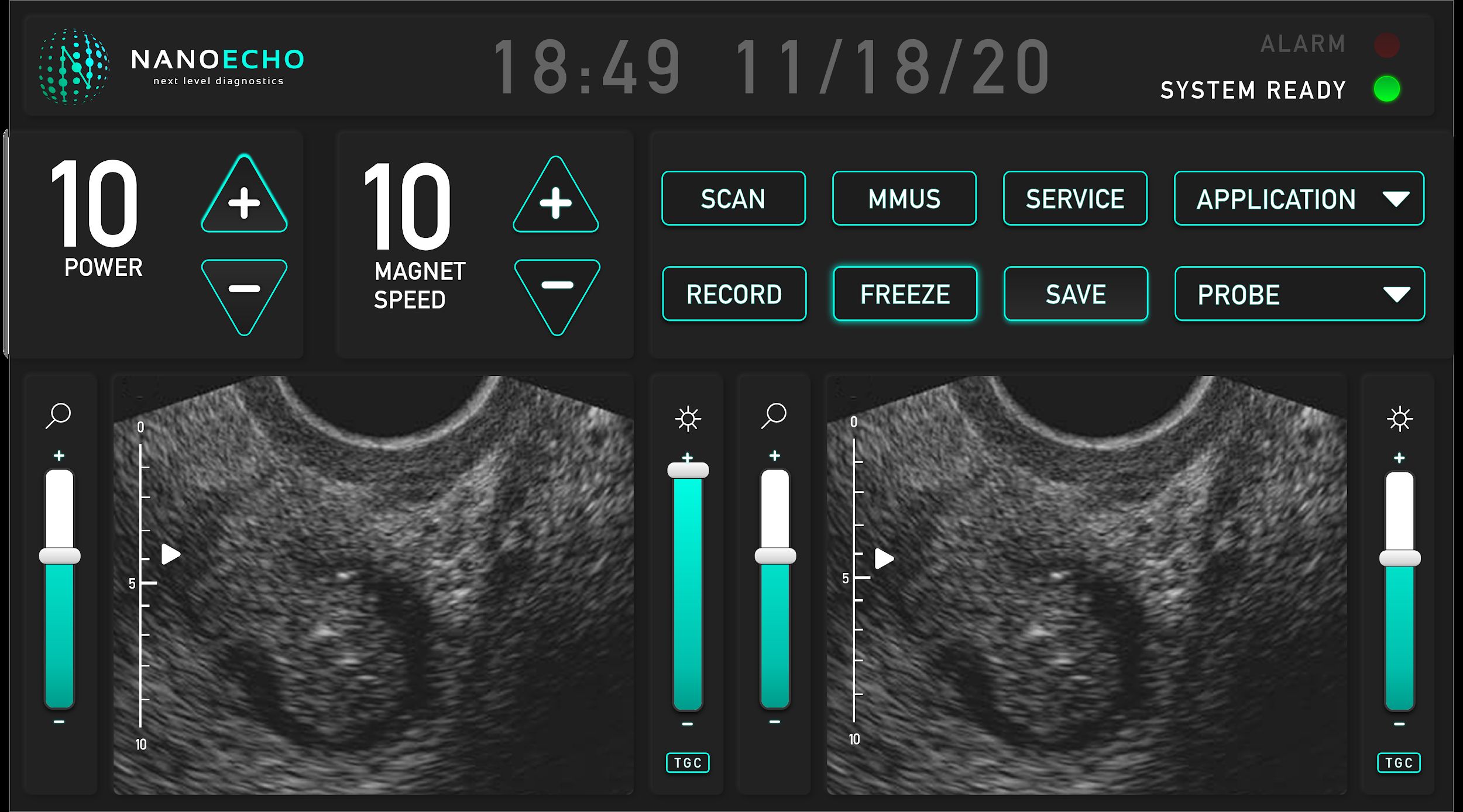
Main scan interface
Live Scan

Dual-view live ultrasound with quick controls for power, magnet, record, freeze, save and probe selection.

Under the hood

A three-tier architecture, designed for the imaging room.

NanoEcho keeps the moving parts cleanly separated so clinical performance never depends on UI responsiveness or device timing. The result is a calm, predictable experience for the clinician — and a maintainable platform for the engineering team.

Device tier

The ultrasonic probe and the magnetic-excitation motor — driven by their native hardware libraries.

Acquisition tier

A Python data-acquisition layer with a state-machine and shared-memory model, exchanging frames over TCP / ZMQ.

Render tier

A real-time 3D engine (Unreal Engine 4) renders the volume — the same fidelity used in modern games, now in a clinical context.

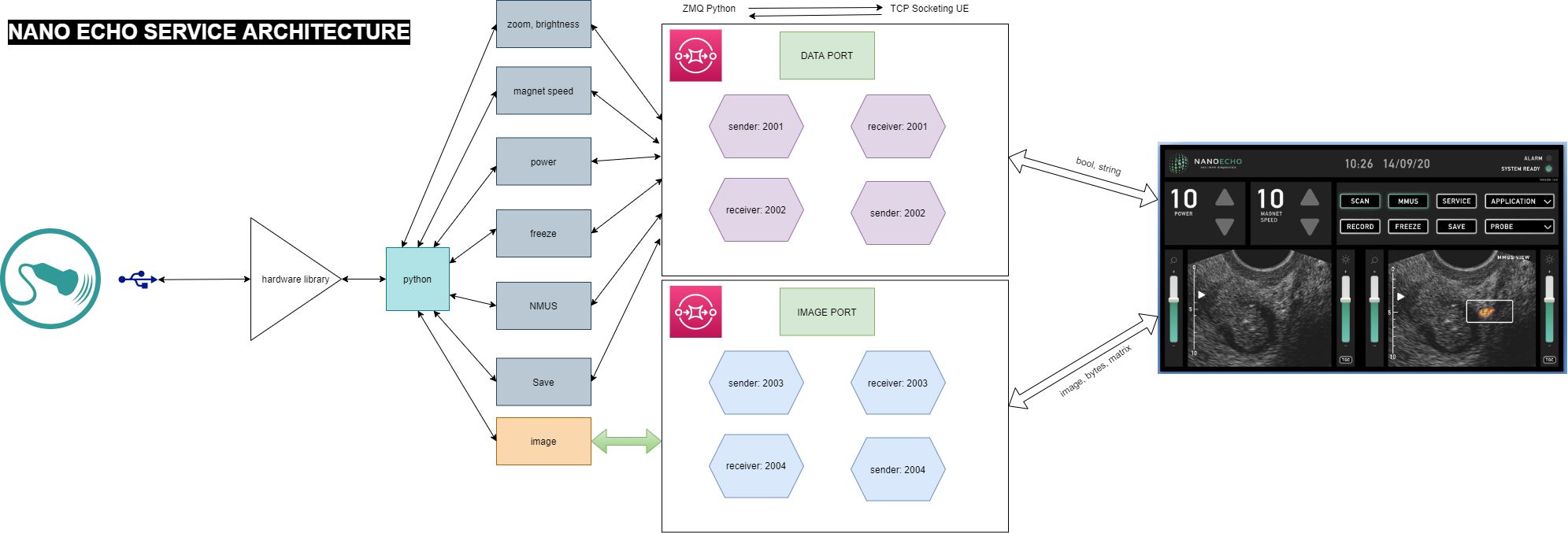
Ultrasonic probe Maxon motor + magnet Python acquisition State machine TCP · ZMQ Computer Vision Unreal Engine 4
NanoEcho three-tier architecture diagram
The three-tier service architecture — devices, Python acquisition, render UI — communicating via ZMQ on the data & image ports.
AI inside

Computer vision does the second look.

A trained vision pipeline runs alongside the live scan — sharpening the image, calling out anomalies, and giving the clinician a head-start.

Auto-highlight findings

Suspicious regions get a soft, non-intrusive overlay — never a final verdict, always a "look here" hint for the human.

Lower-latency imaging

Optimised vision pipelines compress the time from probe-to-pixel, keeping the live view responsive even under heavy scan loads.

Better visualisation

Noise reduction and contrast enhancement make subtle anatomy easier to read — without obscuring the underlying truth.

What it delivers

Better diagnostics, by design.

3D

Volumetric view

Replace 2D slices with a rotatable, sliceable 3D volume — explore anatomy the way clinicians actually think.

AI

Anomaly highlighting

Computer-vision pre-annotates regions of concern, so reading time is spent on judgement, not hunting.

Reduced latency

Tight Python ↔ Unreal pipeline keeps live frames responsive — no "scan now, see later" friction.

Earlier detection

Nano-particle excitation surfaces lesions and inflammations that conventional ultrasound can quietly miss.

Service-grade UI

Separate operator and service modes, always-visible alarm and system-ready states — built for clinical reality.

Extensible probe set

The architecture welcomes new probes, new vision models and new render passes without touching the clinician's workflow.

Bring NanoEcho to your clinical workflow

Ready to give your clinicians a better view?

Whether it's diagnostic imaging, surgical planning, or remote consultation — we can stand up an AI-assisted, real-time 3D imaging platform on top of your hardware.